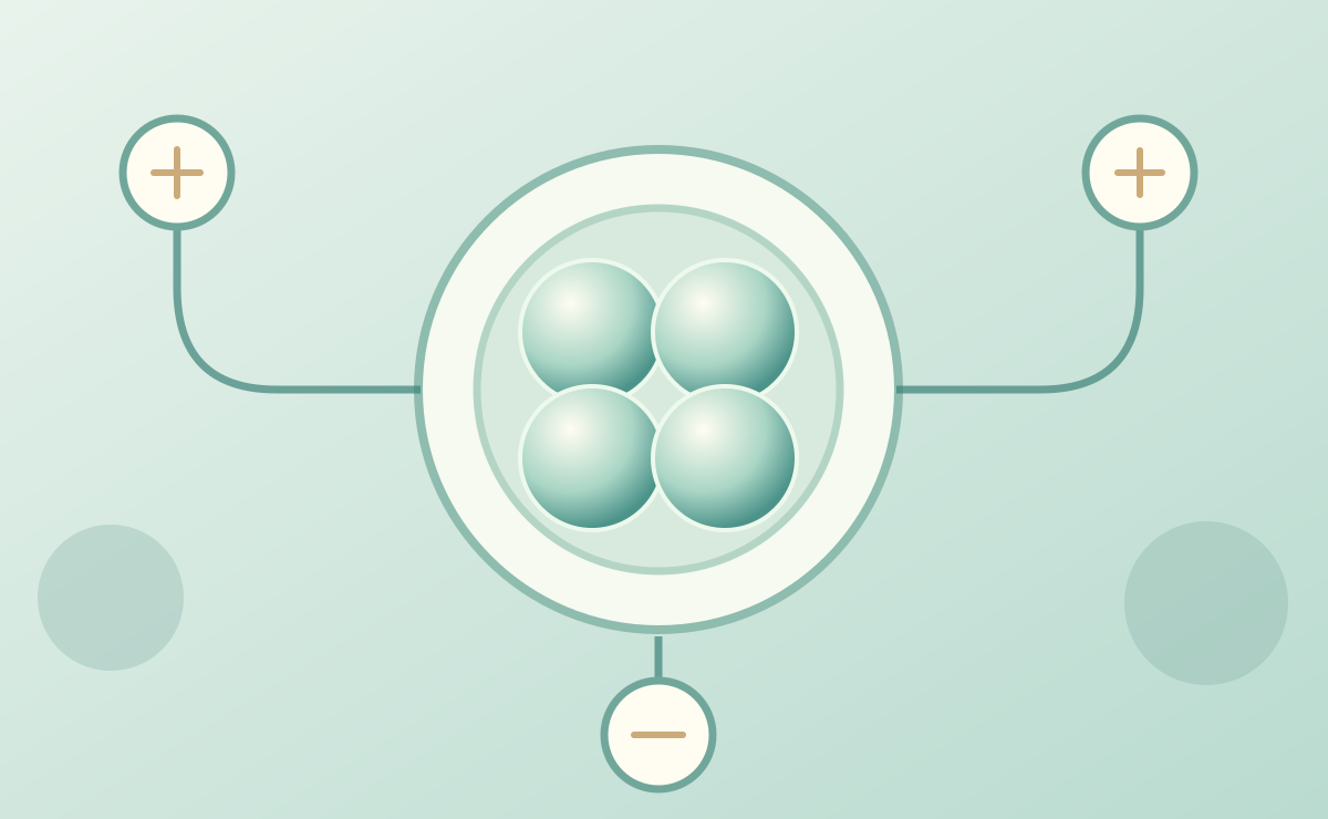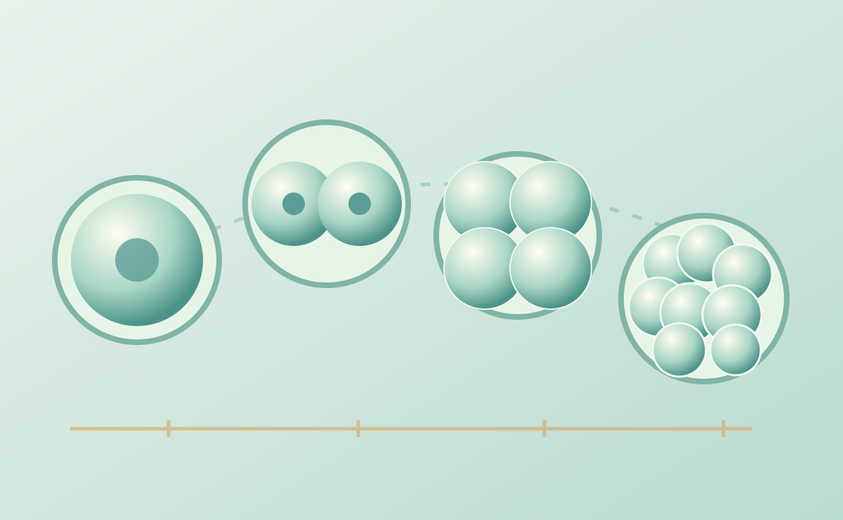
What do the timing and sequence of early cleavage events reveal about developmental processes, beyond a single morphological snapshot?
Why this question matters
Time-lapse imaging transforms embryo assessment from isolated observations into a developmental history. Parameters such as the times of early cleavages, morula formation, and blastulation describe distinct events, but their biological interpretation depends on culture conditions, species, measurement definitions, and study design.
How the question can be studied
This direction integrates longitudinal imaging, consistent event annotation, blastocyst morphology, and cellular or molecular endpoints such as immunofluorescence. The emphasis is on transparent definitions, uncertainty, and validation rather than assuming that any single timing metric predicts an outcome.
What the work may contribute
Linking dynamic observations to independent biological measurements may improve our understanding of early development and identify hypotheses worth testing in larger, independent datasets. Clinical prediction requires separate prospective validation.
Questions still open
- Which morphokinetic relationships are robust across laboratories and imaging platforms?
- How do temporal features relate to cellular phenotypes at the blastocyst stage?
- How can small datasets avoid overfitting and exaggerated predictive claims?
Publications and resources
Related verified publications will be linked here after the bibliography is added.